


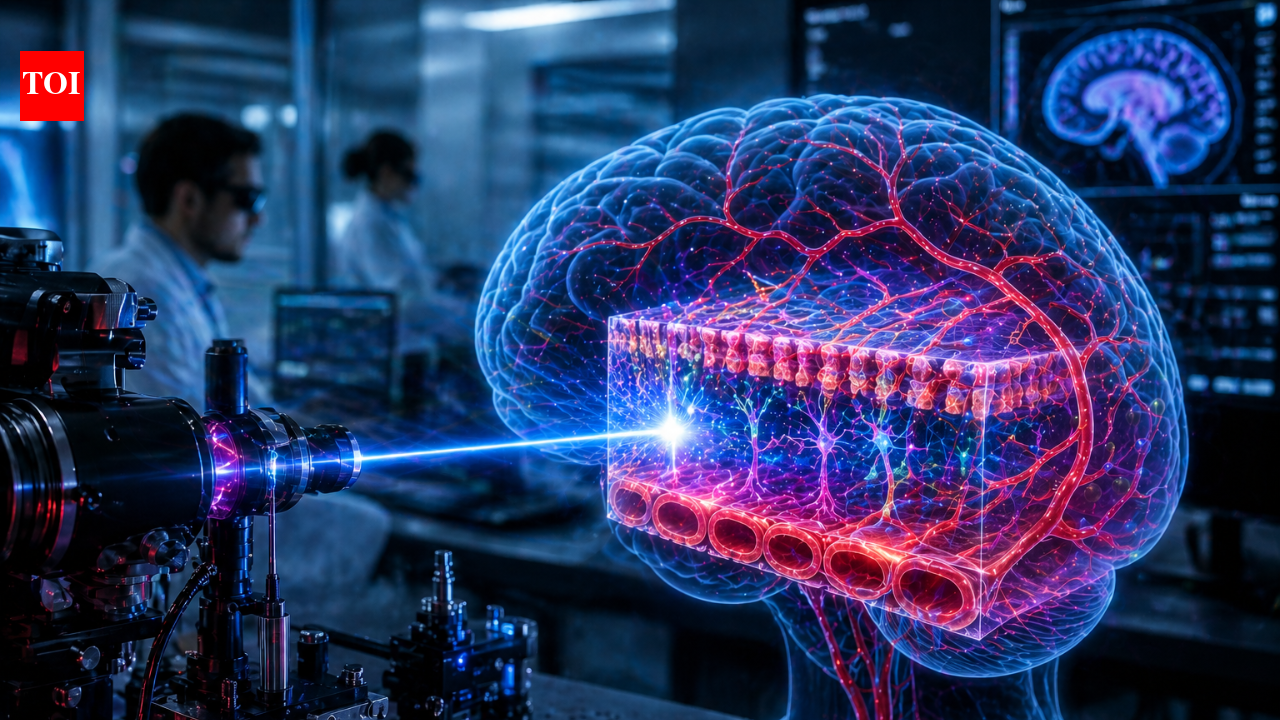
A team of researchers at the Massachusetts Institute of Technology has discovered that chaotic laser light can be transformed into a highly focused imaging tool, enabling scientists to study brain biology at unprecedented speed. The technique converts disordered light travelling through an optical fibre into a stable “pencil beam,” allowing 3D imaging of the blood-brain barrier roughly 25 times faster than existing methods. By capturing cellular activity in real time, the approach could significantly improve how researchers evaluate whether drugs reach the brain, offering a powerful new tool for investigating neurological diseases.
Chaotic laser light becomes a 25 times faster brain imaging tool
The study, published in Nature Methods in April 2026, was led by Sixian You, with lead author Honghao Cao and collaborators from MIT, Harvard University and Beth Israel Deaconess Medical Center.Researchers found that when laser light is sent through a multimode optical fibre under very specific conditions, it does not scatter randomly as expected. Instead, it self-organises into a narrow, intense beam known as a “pencil beam.” This behaviour emerges when the laser is precisely aligned at a near-perfect angle and its power is increased to a level where the light begins interacting with the fibre material itself. At that critical point, nonlinear optical effects counterbalance the inherent disorder inside the fibre, producing a clean and stable beam without the need for complex beam-shaping equipment.
Faster and clearer imaging of the blood-brain barrier
Using this self-organised beam, the team achieved high-resolution, volumetric imaging of the blood-brain barrier, a tightly packed layer of cells that protects the brain from harmful substances.Traditional imaging techniques typically capture one two-dimensional slice at a time, requiring repeated scans to build a full three-dimensional picture. This process limits both speed and the ability to observe dynamic biological processes. In contrast, the new method enables rapid 3D imaging at cellular resolution while maintaining a large depth of focus. It also reduces common artefacts such as blurred halos, resulting in clearer images. Overall, the system delivers imaging speeds approximately 25 times faster than current gold-standard approaches while preserving comparable quality.
Real-time tracking of drugs entering brain cells
One of the most important applications of this technique is its ability to observe how substances move through the blood-brain barrier in real time.This is particularly relevant for conditions such as Alzheimer’s disease and Amyotrophic lateral sclerosis, where drug delivery to the brain remains a major challenge. Many promising treatments fail because they cannot effectively cross this protective barrier.The new imaging approach allows researchers to directly monitor how drugs enter brain cells and measure the rate at which different cell types absorb them. Importantly, it does not require fluorescent labelling, which is commonly used in traditional techniques and can interfere with natural biological processes. This capability could improve how pharmaceutical researchers test therapies using human-based tissue models and better predict how drugs will behave in the human brain.
A simpler approach with broad scientific potential
Another advantage of the method is its relative simplicity. The system works with standard optical components and does not rely on highly specialised beam-shaping tools, making it more accessible to laboratories that may not have advanced optical engineering capabilities.This accessibility could accelerate adoption across multiple scientific fields, including neuroscience, pharmacology and biomedical engineering. At the same time, the discovery challenges long-standing assumptions in optical physics, where disorder in laser systems has traditionally been viewed as a limitation rather than a potential advantage.
what comes next for the technology
The team plans to further investigate the physical mechanisms behind this self-organising behaviour and explore additional applications, including imaging neurons and other complex biological systems.While the technique remains at an early research stage, it represents a significant step forward in bioimaging. By transforming chaotic laser light into a precise and efficient imaging tool, scientists have opened new possibilities for studying the brain and advancing research into neurological diseases.